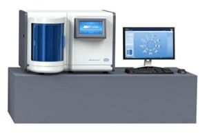
In this study, we compared Ki-67 LI between by VA and by ADIA, and analyzed the causes of discrepancies. VA and ADIA were shown have their own strengths and weaknesses. We performed visual assessment (VA) and ADIA simultaneously for breast carcinoma cases for approximately seven months. Several studies have observed ADIA to yield more reproducible and accurate measurement, and its application is being implemented in a clinical setting. Automated digital image analysis (ADIA) of Ki-67 LI in breast cancers obtains high quality data. Recently, computer-assisted image analysis has been used to achieve higher reproducibility of IHC results. Several methods, such as a five-grade scale, have been suggested to resolve this problem however, low reproducibility remains an issue.

Despite its importance, Ki-67 LI has high inter-observer and/or intra-observer variability, and low reproducibility.
Traditionally, Ki-67 LI is estimated by visual observation. Recurrence rate, prognosis, and therapeutic recommendations differ according to subtypes. For instance, in ER-positive and HER2-negative breast cancers in particular, the classification of subtypes is dependent on Ki-67 LI: tumors with low Ki-67 LI are classified into the luminal A group and those with high Ki-67 LI into the luminal B group. Among them, Ki-67 LI is a parameter for molecular classification and prognostic assessment. Immunohistochemistry (IHC) is widely used to determine the expression of these markers. Currently, the molecular classification of breast carcinomas can be easily confirmed according to estrogen receptor (ER), progesterone receptor (PR), human epidermal growth factor 2 (HER2), or Ki-67 labeling index (LI) status. Intensive molecular studies have made it possible to classify breast carcinomas, leading to improvements in treatment, prognosis prediction, and outcomes. Many studies have sought to improve treatment outcomes for breast cancer, and molecular studies play a critical role in prognosis. Therefore, ADIA findings require confirmation by a pathologist.īreast cancer is one of the most frequent malignancies in women. Although ADIA is more accurate than VA, there are some limitations. There were more discrepancies between VA and ADIA results in the group with a VA value of 10–20% compared to groups with <10% and ≥20%.

The cause of these differences was: (1) tumor heterogeneity (98 cases, 56.0%), (2) VA interpretation error (32 cases, 18.3%), (3) misidentification of tumor cells (26 cases, 14.9%), (4) poor immunostaining or slide quality (16 cases, 9.1%), and (5) Estimation of non-tumor cells (3 cases, 1.7%). We retrospectively analyzed cases with a greater than 5% difference between VA and ADIA results. VA and ADIA were highly correlated (intraclass correlation coefficient 0.982, and Spearman’s correlation coefficient 0.966, p<0.05). We used both VA and automated digital image analysis (ADIA) (Ventana Virtuoso image management software) to estimate Ki-67 LI for 997 cases of breast carcinoma, and compared VA and ADIA results. For more accurate assessment and better reproducibility with Ki-67 LI, digital image analysis was introduced recently. The Ki-67 LI is traditionally calculated via unaided microscopic estimation however, inter-observer and intra-observer variability and low reproducibility are problems with this visual assessment (VA) method. The Ki-67 labeling index (LI) is an important prognostic factor in breast carcinoma.


 0 kommentar(er)
